Plantar Fascia Surgery
Take the first step to better health with Dr. Chowdhury, our highly experienced Foot & Ankle Surgeon!
Request Appointment

Overview
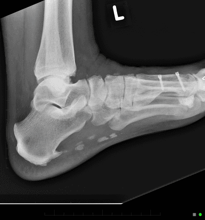
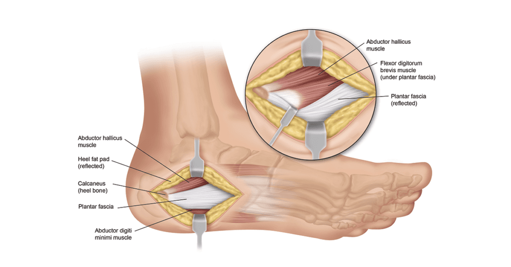
Plantar fasciitis is an inflammatory condition caused by tearing of the plantar fascia, or plantar aponeurosis. Often, it arises as a secondary issue due to a tight Achilles tendon, which pulls on the heel bone, resulting in tension on the plantar fascia. This tearing leads to inflammation, pain, and swelling. In chronic cases, the body may attempt to alleviate tension by elongating the heel bone, forming a heel spur. Contrary to common belief, the pain does not originate from the heel spur but rather from micro-tears in the fascia and the ensuing inflammation.
Request Appointment
In Need of Plantar Fascia Surgery?
Symptoms
Patients with plantar fasciitis typically experience sharp pain in the heel, often described as a stabbing sensation, particularly noticeable during the first steps in the morning or after prolonged periods of rest. While the pain tends to lessen with activity, it can persist throughout the day in chronic cases.
Diagnosis
A thorough clinical history and physical examination are usually sufficient for diagnosis. Palpation of the medial calcaneal tubercle is a common finding, along with limitations in ankle range of motion. Another indicator is the inability to move the little toe outward.
Diagnostic Methods
Various imaging studies, including X-rays, MRI, and ultrasound, aid in assessing plantar fasciitis. X-rays can detect heel spurs and rule out other conditions, while MRI provides detailed visualization of soft tissue structures and inflammation. Ultrasound offers real-time visualization and is useful for guided diagnostic injections.
When to Seek Medical Advice
Medical attention is warranted if conservative measures like rest, icing, stretching, and over-the-counter inserts fail to alleviate symptoms, or if symptoms worsen. Changes in foot anatomy, persistent pain, or mobility limitations also necessitate evaluation.
Treatment Options
Non-Surgical Treatments:
Shockwave therapy and consistent stretching, guided by a physical therapist, are effective non-operative treatments. Shockwave therapy promotes tissue healing and reduces pain, typically requiring 3-6 sessions.
Surgical Options:
EPF involves cutting a portion of the plantar fascia using a camera and cutting instrument. Plantar fasciectomy with bone spur removal is a mini-open procedure where the problematic portion of the fascia is removed. Gastrocnemius recession indirectly relieves tension on the plantar fascia by cutting a thin, soft tissue structure overlying the calf muscle.
Rehabilitation & Recovery
Post-operative care involves minimal downtime, with all procedures performed on an outpatient basis. Patients can typically resume walking on the day of surgery with the aid of a special shoe or boot. Recovery involves keeping the surgical dressing clean and dry, elevating the foot, and taking pain medication as needed. Gradual return to activities is expected within a few weeks, sometimes supplemented by physical therapy for optimal rehabilitation.


Plantar Fascia Surgery
Take the first step to better health with Dr. Chowdhury, our highly experienced Foot & Ankle Surgeon!
Plantar Fascia Surgery
Take the first step to better health with Dr. Chowdhury, our highly experienced Foot & Ankle Surgeon!


Overview
Plantar fasciitis is an inflammatory condition caused by tearing of the plantar fascia, or plantar aponeurosis. Often, it arises as a secondary issue due to a tight Achilles tendon, which pulls on the heel bone, resulting in tension on the plantar fascia. This tearing leads to inflammation, pain, and swelling. In chronic cases, the body may attempt to alleviate tension by elongating the heel bone, forming a heel spur. Contrary to common belief, the pain does not originate from the heel spur but rather from micro-tears in the fascia and the ensuing inflammation.
Diagnosis
A thorough clinical history and physical examination are usually sufficient for diagnosis. Palpation of the medial calcaneal tubercle is a common finding, along with limitations in ankle range of motion. Another indicator is the inability to move the little toe outward.
Symptoms
Patients with plantar fasciitis typically experience sharp pain in the heel, often described as a stabbing sensation, particularly noticeable during the first steps in the morning or after prolonged periods of rest. While the pain tends to lessen with activity, it can persist throughout the day in chronic cases.
Treatment Options
Non-Surgical Treatments:
Shockwave therapy and consistent stretching, guided by a physical therapist, are effective non-operative treatments. Shockwave therapy promotes tissue healing and reduces pain, typically requiring 3-6 sessions.
Surgical Options:
EPF involves cutting a portion of the plantar fascia using a camera and cutting instrument. Plantar fasciectomy with bone spur removal is a mini-open procedure where the problematic portion of the fascia is removed. Gastrocnemius recession indirectly relieves tension on the plantar fascia by cutting a thin, soft tissue structure overlying the calf muscle.
Diagnostic Methods
Various imaging studies, including X-rays, MRI, and ultrasound, aid in assessing plantar fasciitis. X-rays can detect heel spurs and rule out other conditions, while MRI provides detailed visualization of soft tissue structures and inflammation. Ultrasound offers real-time visualization and is useful for guided diagnostic injections.
When to Seek Medical Advice
Medical attention is warranted if conservative measures like rest, icing, stretching, and over-the-counter inserts fail to alleviate symptoms, or if symptoms worsen. Changes in foot anatomy, persistent pain, or mobility limitations also necessitate evaluation.
Rehabilitation & Recovery
Post-operative care involves minimal downtime, with all procedures performed on an outpatient basis. Patients can typically resume walking on the day of surgery with the aid of a special shoe or boot. Recovery involves keeping the surgical dressing clean and dry, elevating the foot, and taking pain medication as needed. Gradual return to activities is expected within a few weeks, sometimes supplemented by physical therapy for optimal rehabilitation.


Plantar Fascia Surgery
Take the first step to better health with Dr. Chowdhury, our highly experienced Foot & Ankle Surgeon!
Plantar Fascia Surgery
Take the first step to better health with Dr. Chowdhury, our highly experienced Foot & Ankle Surgeon!


SPORTS FOOT &
ANKLE CENTER
Services
Achilles Tendonitis
Ankle Fracture
Lisfranc Injury
Ankle Sprain
... + 20 more
Reviews
Jessica Peri
Sameer Alam
Noman Saleemi
Andres Botero
…+ 6 more
Contact
201-777-1245
dr.einfootandankle@gmail.com
Location
SPORTS FOOT &
ANKLE CENTER
Services
Achilles Tendonitis
Ankle Fracture
Lisfranc Injury
Ankle Sprain
... + 20 more
Reviews
Jessica Peri
Sameer Alam
Noman Saleemi
Andres Botero
…+ 6 more
Location
Contact
201-777-1245
dr.einfootandankle@gmail.com


Request Appointment
In Need of Plantar Fascia Surgery?
SPORTS FOOT &
ANKLE CENTER
Services
Achilles Tendonitis
Ankle Fracture
Lisfranc Injury
Ankle Sprain
... + 20 more
Testimonials
Jessica Peri
Sameer Alam
Noman Saleemi
Andres Botero
…+ 6 more
Location
Contact
201-777-1245
dr.einfootandankle
@gmail.com


Overview
Plantar fasciitis is an inflammatory condition caused by tearing of the plantar fascia, or plantar aponeurosis. Often, it arises as a secondary issue due to a tight Achilles tendon, which pulls on the heel bone, resulting in tension on the plantar fascia. This tearing leads to inflammation, pain, and swelling. In chronic cases, the body may attempt to alleviate tension by elongating the heel bone, forming a heel spur. Contrary to common belief, the pain does not originate from the heel spur but rather from micro-tears in the fascia and the ensuing inflammation.
Treatment Options
Non-Surgical Treatments:
Shockwave therapy and consistent stretching, guided by a physical therapist, are effective non-operative treatments. Shockwave therapy promotes tissue healing and reduces pain, typically requiring 3-6 sessions.
Surgical Options:
EPF involves cutting a portion of the plantar fascia using a camera and cutting instrument. Plantar fasciectomy with bone spur removal is a mini-open procedure where the problematic portion of the fascia is removed. Gastrocnemius recession indirectly relieves tension on the plantar fascia by cutting a thin, soft tissue structure overlying the calf muscle.
When to Seek Medical Advice
Medical attention is warranted if conservative measures like rest, icing, stretching, and over-the-counter inserts fail to alleviate symptoms, or if symptoms worsen. Changes in foot anatomy, persistent pain, or mobility limitations also necessitate evaluation.
Diagnostic Methods
Various imaging studies, including X-rays, MRI, and ultrasound, aid in assessing plantar fasciitis. X-rays can detect heel spurs and rule out other conditions, while MRI provides detailed visualization of soft tissue structures and inflammation. Ultrasound offers real-time visualization and is useful for guided diagnostic injections.
Symptoms
Patients with plantar fasciitis typically experience sharp pain in the heel, often described as a stabbing sensation, particularly noticeable during the first steps in the morning or after prolonged periods of rest. While the pain tends to lessen with activity, it can persist throughout the day in chronic cases.
Diagnosis
A thorough clinical history and physical examination are usually sufficient for diagnosis. Palpation of the medial calcaneal tubercle is a common finding, along with limitations in ankle range of motion. Another indicator is the inability to move the little toe outward.
Rehabilitation & Recovery
Post-operative care involves minimal downtime, with all procedures performed on an outpatient basis. Patients can typically resume walking on the day of surgery with the aid of a special shoe or boot. Recovery involves keeping the surgical dressing clean and dry, elevating the foot, and taking pain medication as needed. Gradual return to activities is expected within a few weeks, sometimes supplemented by physical therapy for optimal rehabilitation.